
Information
History
The Department of Clinical Oncology at The Chinese University of Hong Kong/ Prince of Wales Hospital is an integrated academic and clinical unit providing state-of-the-art cancer care to a catchment population of 1.8 million residents within the New Territories East Cluster (NTEC) of Hong Kong, as well as a referral centre for tertiary/quaternary cancer care for the entire population of Hong Kong as a whole. We are a high-volume cancer centre with an average 4000 new patients being seen yearly.
We provide quality cancer care ranging from active treatment, to palliative and psychosocial care and hospice services in both inpatient and outpatient settings, through a multi-disciplinary approach, by a dedicated team of medical oncologists, haematological oncologists, radiation oncologists/clinical oncologists, palliative medicine specialists, nurses, radiation therapists, medical physicists, clinical pharmacists and other allied health professionals.
Academic Interests
Our academic interests are on clinical and traditional researches for lung cancer, nasopharyngeal cancer, head & neck cancer, sarcoma/melanoma, genitourinary cancer, haematological, gastrointestinal & hepatocellular cancers.
Current Trials
The Comprehensive Cancer Trials Unit (CCTU) have conducted over 100
clinical and translational studies to date, with active collaborations both regionally and
internationally.


Research Postgraduate
Research in the Tumour Marker Laboratory involves two main areas: screening of novel anticancer drugs
and the study.

Events
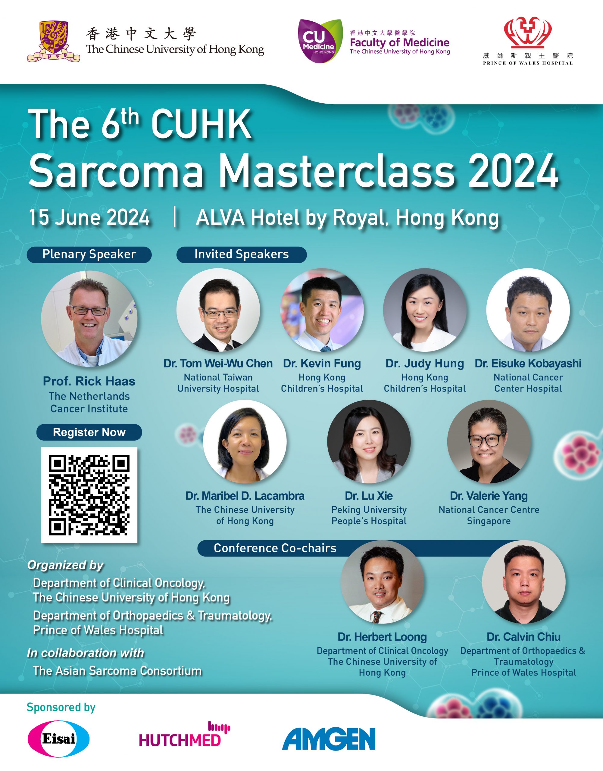
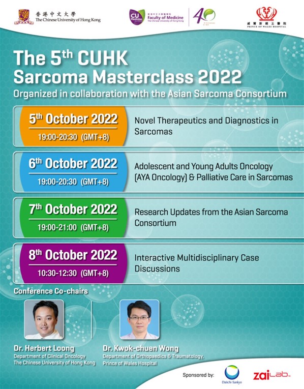
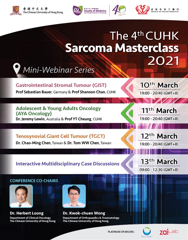
The 6th CUHK Sarcoma Masterclass 2024


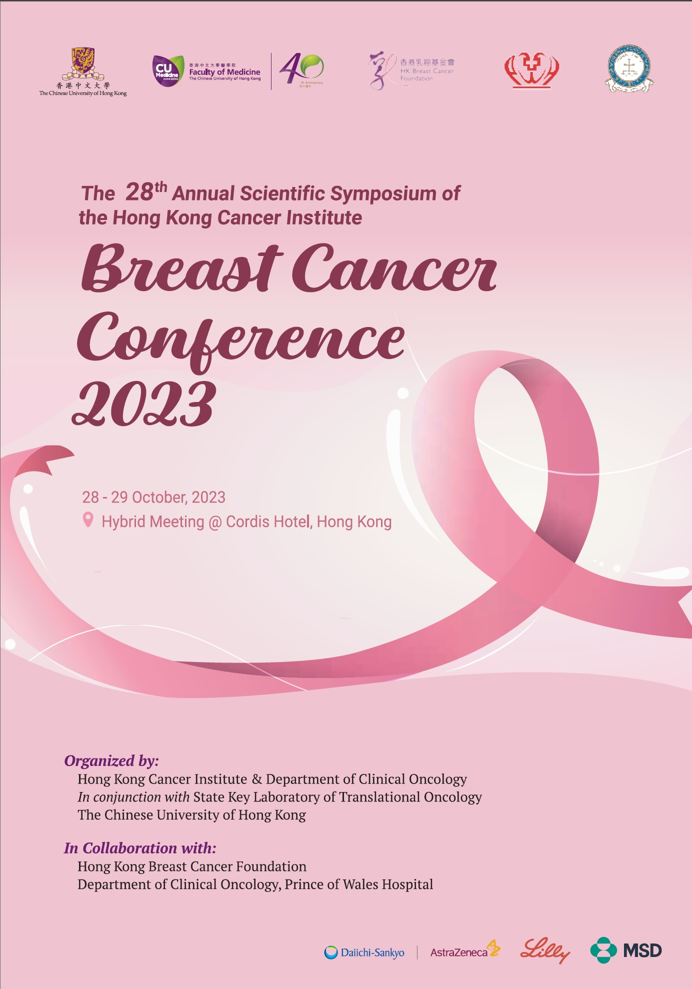
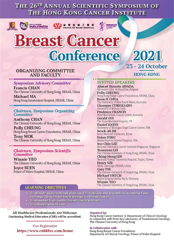
Breast Cancer Conference 2023
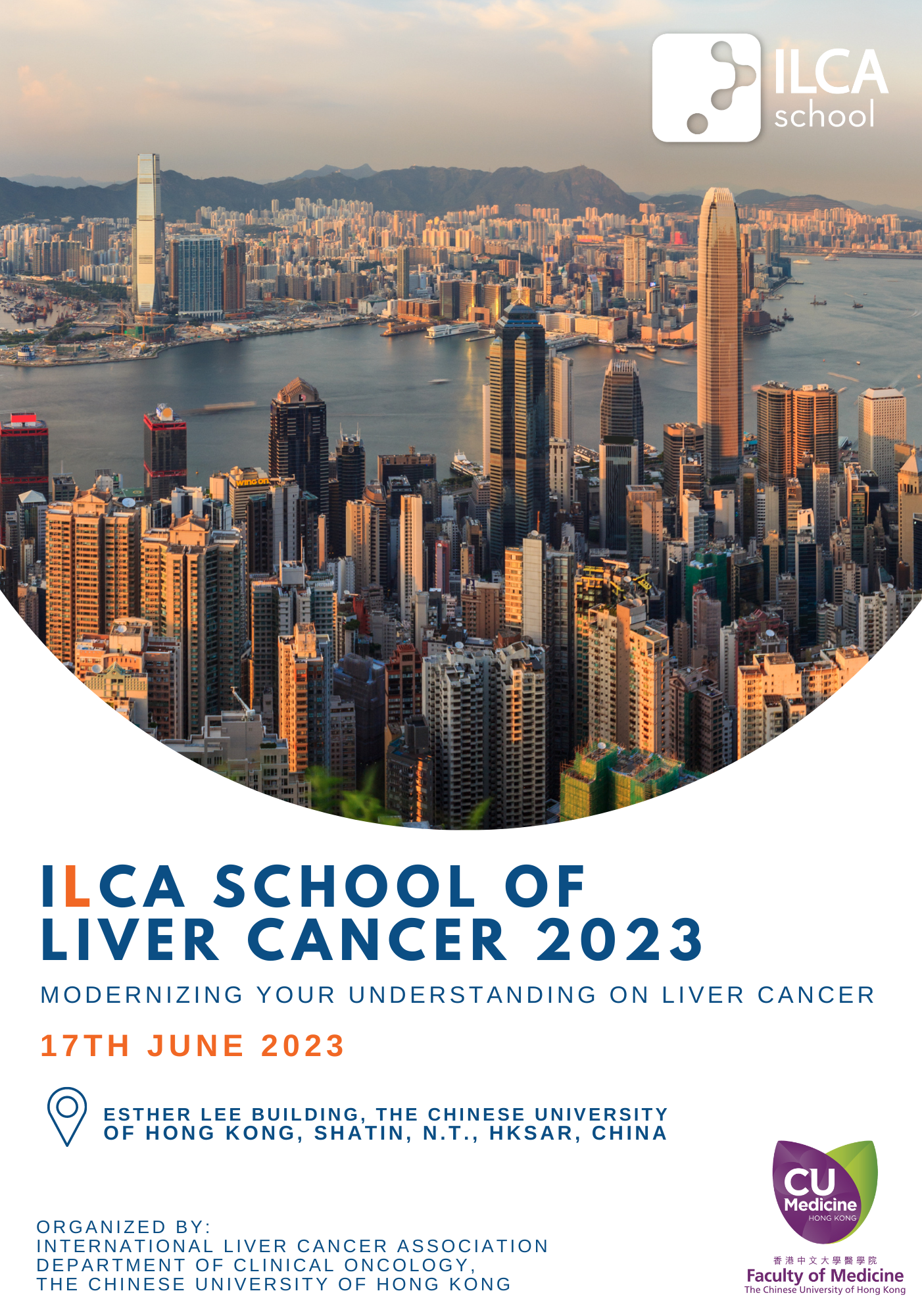
ILCA School of Liver Cancer 2023
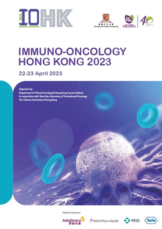
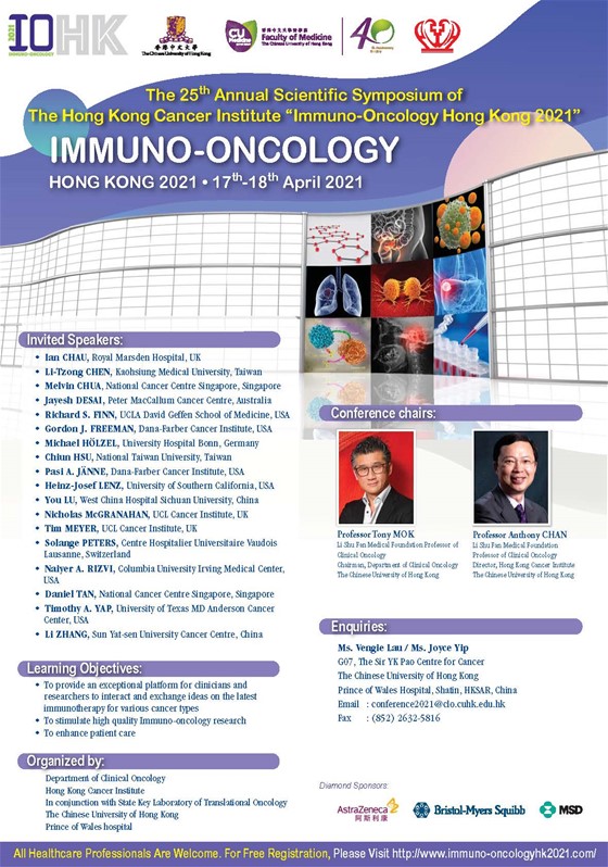
Immuno-Oncology Hong Kong 2023
Publications
Li CH, To KF, Tong JH, Xiao Z, Xia T, Lai PB, Chow SC, Zhu YX, Chan SL, Marquez VE, Chen Y. Enhancer of zeste homolog 2 silences mir-218 in human pancreatic ductal adenocarcinoma cells by inducing formation of heterochromatin. Gastroenterology 2013; 144:1086-1097. [PubMed]
Mesia R, Henke M, Fortin A, Minn H, Yunes Ancona AC, Cmelak A, Markowitz AB, Hotte SJ, Singh S, Chan AT, Merlano MC, Skladodwski K, Zhang A, Oliner KS, VanderWalde A, Giralt J. Chemmradiotherapy with or without panitumumab in patients with unresected, locally advanced squamous-cell carcinoma of the head and neck (CONCERT-1): a randomized, controlled, open-label phase 2 trial. Lancet Oncol 2015; 16(2):208-20. [PubMed]
Ribassin-Majed L., Marguet S., Lee A.W., Ng W.T., Ma J., Chan A.T., Huang P.Y., Zhu G., Chua D.T., Chen Y., Mai H.Q., Kwong D.L., Cheah S.L., Moon J., Tung Y., Chi K.H., Fountzilas G., Bourhis J., Pignon J.P., Blanchard P. What is the best treatment of locally advanced Nasopharyngeal Carcinoma? An individual patient data network meta-analysis. J Clin Oncol 2016 Dec 5 (Epub) [PubMed]


